

The subluxation meaning varies significantly depending on the clinical context in which it is applied. At its core, the term describes a partial or incomplete dislocation of a joint, where the bony surfaces remain in partial contact with one another. Unlike a complete dislocation—where the joint surfaces are entirely separated—a subluxation represents a state of mechanical instability that can range from asymptomatic to severely debilitating.
Because the term is used across distinct disciplines, including orthopedics, dentistry, and chiropractic care, patients often encounter confusion when attempting to interpret diagnostic findings. Gaining clarity on these nuances is essential for understanding how different practitioners assess musculoskeletal and structural integrity.
Orthopedic and General Medical Definitions
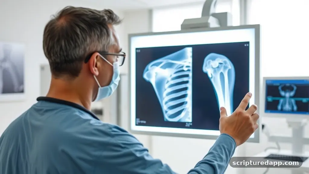
In the realm of general medicine and orthopedics, the subluxation meaning is strictly anatomical. It refers to a joint that has lost its normal alignment, yet the articular surfaces remain partially opposed.
When a trauma occurs, the ligaments and capsule surrounding the joint may be stretched or torn, allowing the bones to shift out of their natural position. This is distinct from a full dislocation, where the joint surfaces lose all contact, often requiring physical manipulation or surgical intervention to restore alignment.
Clinicians typically diagnose this condition through physical examination and diagnostic imaging, such as X-rays or MRI scans. The clinical significance of a subluxation often depends on the joint involved. For example, a shoulder subluxation is a common occurrence in athletes or individuals with hypermobility, leading to recurrent episodes of instability and discomfort.
In these cases, the joint may temporarily slip out of place and then shift back on its own. The underlying trauma often compromises the soft tissue support, creating a cycle where the joint becomes increasingly prone to further displacement. Treatment generally focuses on stabilizing the joint through physical therapy, strengthening the surrounding musculature, and, in severe cases, surgical stabilization to prevent future occurrences.
The Chiropractic Perspective on Vertebral Subluxation
Within the chiropractic profession, the vertebral subluxation is a foundational concept, though its definition differs from the strictly anatomical view used in conventional orthopedics. Chiropractors often describe this as a complex of functional, structural, and neurological changes that compromise the integrity of the spine. Rather than focusing solely on the displacement of bone, this view emphasizes how the altered position or movement of a vertebra impacts the nervous system.
Practitioners who focus on this model argue that when a vertebra is misaligned or experiencing restricted movement, it can interfere with the transmission of neural signals. This interference is thought to produce various symptoms that affect overall health. While traditional medicine often requires objective, radiographically visible displacement to define a subluxation, the chiropractic model includes “fixations” or “motion restrictions” that may not be apparent on standard imaging.
This has historically led to a divergence in terminology and application. Proponents of this approach believe that by performing spinal adjustments to correct these subluxations, they can improve joint mobility, reduce nerve irritation, and restore the body’s natural ability to function at its peak. This practice has been a cornerstone of the field for over a century, evolving as clinical research and patient outcomes continue to inform the way practitioners approach spinal health.
Clinical Differences: Subluxation vs. Dislocation
Understanding the distinction between a subluxation and a complete dislocation is critical for effective patient communication. While both conditions represent a disruption of joint integrity, the severity and immediate management requirements differ.
A dislocation is an acute, high-impact injury where the joint is “locked” or rendered immobile because the articular surfaces have completely moved past one another. The surrounding tissues are often significantly damaged, and the patient experiences intense pain and a visible deformity of the affected area.
Conversely, a subluxation is often more subtle. The joint may exhibit a “giving way” sensation or a feeling of looseness.
Because the surfaces remain partially in contact, the joint may technically remain functional, albeit compromised. The following table highlights the primary differences between these two conditions:
| Feature | Subluxation | Dislocation |
|---|---|---|
| Joint Contact | Partial contact remains | Complete loss of contact |
| Visual Appearance | Often subtle or normal | Obvious deformity |
| Mobility | Reduced or unstable | Locked or immobile |
| Urgency | Requires evaluation/therapy | Emergency medical reduction |
Recognizing these nuances is vital. A patient experiencing a subluxation might not seek immediate care, assuming the issue is minor, while a dislocation is almost universally treated as an urgent medical event. However, repeated subluxations can lead to chronic joint degradation, often referred to as “joint laxity,” which necessitates proactive intervention to prevent long-term disorders.
Dental and Oral Health Subluxations
The subluxation meaning also extends into the field of dentistry, specifically regarding the temporomandibular joint (TMJ) and dental trauma. In a dental context, a subluxation of a tooth occurs when the tooth is loosened in its socket due to impact, but it is not completely avulsed (knocked out).
The periodontal ligament is damaged, causing the tooth to become mobile or displaced within the alveolar bone. This is a common dental emergency that requires immediate stabilization to ensure the tooth can reattach and remain viable.
Furthermore, TMJ subluxation occurs when the jaw joint moves beyond its normal range of motion. This often manifests as a clicking or popping sound when opening the mouth, or in more severe cases, the jaw may become temporarily locked in an open position. Patients suffering from this condition often experience pain and difficulty chewing.
Dentists and oral surgeons treat these cases by assessing the occlusion (how the teeth meet) and the integrity of the TMJ disc. Treatment might involve night guards, physical therapy to strengthen the muscles around the jaw, or, if the joint capsule is chronically loose, surgical interventions to tighten the ligaments and prevent the jaw from slipping out of the socket during normal function.
Historical Evolution of the Term
The history of the term subluxation is as varied as the fields that utilize it. Etymologically, it is derived from the Latin words “sub” (meaning beneath or less than) and “luxare” (meaning to dislocate).
Historically, the term was used more broadly in early medical literature to describe any joint that was not perfectly aligned. As medical imaging techniques like radiography evolved, the definition became more precise in orthopedic circles, requiring visible, objective evidence of misalignment for the diagnosis to be valid.
In the late 19th and early 20th centuries, as the chiropractic profession was created, the term was adopted to describe the specific spinal issues that practitioners identified as the source of patient health complaints. This created a lasting tension between the medical definition—which relies on clear, physical displacement—and the functional definition used by many manual therapists.
Over time, the science behind these concepts has been scrutinized, leading to increased efforts to bridge the gap through modern clinical research. Today, the term continues to evolve as practitioners use advanced technology to study the complex biomechanics of the spine, seeking to better understand how movement patterns influence the effects of health on the nervous system.
The Role of Trauma in Joint Instability

Trauma is the most frequent cause of both orthopedic subluxations and dental displacements. Whether it is a high-velocity impact from a car accident or a repetitive stress injury from poor posture, the result is an overload of the stabilizing structures of the joint.
When a joint is subjected to forces beyond its physiological limit, the ligaments—which are responsible for passive stability—become stretched. Once these ligaments lose their elastic recoil, the joint can no longer maintain its center of rotation.
Even minor, chronic trauma can have cumulative effects. For instance, in the spine, micro-trauma from sedentary desk work or improper lifting techniques can lead to what some call “vertebral subluxation complexes.” While not always an acute injury, the cumulative stress on the joints can lead to inflammation and muscle guarding, where the body attempts to splint the area to prevent further damage.
This guarding can lead to secondary issues, including reduced range of motion and chronic discomfort. Understanding that trauma exists on a spectrum—from the macro-trauma of a fall to the micro-trauma of daily habit—is essential for any patient looking to recover from or prevent these issues. You can find more information on joint anatomy and injury prevention through the National Institute of Arthritis and Musculoskeletal and Skin Diseases.
Diagnostic Modalities and Modern Assessment
Modern medicine utilizes a variety of tools to interpret the subluxation meaning in a clinical setting. While a physical exam is the first step, imaging provides the objective data necessary to confirm the structural state of a joint.
X-rays are the gold standard for viewing bone alignment; they allow practitioners to observe the relationship between vertebrae or the position of a humeral head in the glenoid fossa. However, X-rays are static images and may not capture the instability that occurs during movement.
Dynamic imaging, such as videofluoroscopy or stress X-rays, can be used to observe how a joint behaves under load or during motion. This is particularly useful in cases where the patient reports symptoms of instability that are not present while lying still. Additionally, MRI technology is invaluable for evaluating the soft tissues, such as the labrum in the shoulder or the discs in the spine.
By visualizing the ligaments, tendons, and cartilage, clinicians can determine if the subluxation is a result of structural damage or simply muscle imbalance. Advanced diagnostic techniques allow for more personalized treatment plans, ensuring that interventions are targeted toward the specific tissue or mechanical failure causing the patient’s complaints.
Addressing Topical Gaps in Current Literature
A common gap in current literature is the lack of a unified definition that satisfies both the physiological and functional models of joint health. Many patients search for the subluxation meaning only to find conflicting information that labels the term as either “purely medical” or “purely alternative.” This binary view ignores the reality that joints are complex, living systems that possess both structural and functional requirements.
One overlooked aspect is the role of proprioception—the body’s ability to sense its position in space. When a subluxation occurs, the mechanoreceptors in the joint capsule and surrounding muscles are often compromised. This leads to faulty signaling to the brain, which can cause the body to adopt compensatory movement patterns.
These patterns, if left unaddressed, can lead to secondary disorders in other parts of the body. Another gap is the focus on psychological factors. Chronic joint instability can lead to a fear of movement (kinesiophobia), which further exacerbates the condition by leading to muscle atrophy.
By addressing the psychological response to instability, in addition to the mechanical alignment, practitioners can achieve better, more lasting results. This holistic view is increasingly being adopted in modern rehabilitative medicine.
Common Symptoms and Warning Signs
Identifying the signs of a potential subluxation can help patients seek early intervention, potentially preventing the development of chronic issues. While the specific symptoms depend on the joint involved, there are common markers that indicate joint instability. These include:
- Localized pain that fluctuates in intensity based on activity levels.
- A sensation of the joint “popping,” “clicking,” or feeling like it is shifting out of place.
- Reduced range of motion, often described as a feeling of stiffness or “locking.”
- Muscle spasms in the area surrounding the joint, as the body attempts to stabilize the region.
- Numbness, tingling, or radiating pain, particularly if the subluxation is in the spine and affecting nerve pathways.
- A feeling of weakness or instability when performing everyday movements.
These indicators should not be ignored. While some minor joint issues resolve with rest, persistent symptoms suggest that the structural integrity of the joint has been compromised. Early assessment by a healthcare professional can help differentiate between a temporary irritation and a more serious condition that requires targeted treatment, such as physical therapy or specific mobilization techniques.
Comprehensive Treatment Approaches
Effective management of subluxations is rarely achieved through a single intervention. Instead, it requires a multifaceted approach that addresses the structural, muscular, and neurological components of the injury. The first phase of treatment is typically stabilization.
This might involve bracing or taping to support the joint while allowing the damaged ligaments time to heal. Reducing inflammation through rest, ice, or anti-inflammatory medications is also common in the acute phase.
Once the initial inflammation has subsided, the focus shifts to restoring function. This is where physical therapy and chiropractic care overlap significantly. Therapeutic exercises are designed to strengthen the stabilizing muscles that surround the joint.
For example, in a shoulder subluxation, strengthening the rotator cuff is essential for keeping the humeral head centered. In the spine, core stabilization exercises are used to support the vertebral column, reducing the reliance on the ligaments to maintain alignment.
In some cases, manual therapy is used to improve joint mobility, ensuring that the joint moves through its full range without restriction. By combining passive care (like adjustments or massage) with active care (like rehabilitative exercise), patients can often restore normal function and prevent the recurrence of the subluxation.
FAQ: Understanding Joint Subluxation
Is a subluxation the same as a bone being out of place?
A subluxation is a partial dislocation where the joint surfaces remain in partial contact. While the bone is technically “out of its ideal position,” it is not completely separated from the joint, which is the primary difference between a subluxation and a full dislocation.
Can chiropractic adjustments fix a subluxation?
Chiropractors use adjustments to address vertebral subluxations by restoring motion to restricted joints. The goal is to improve joint function, reduce muscle tension, and alleviate nerve irritation, thereby allowing the body to function more efficiently and support the healing of the surrounding tissues.
Are subluxations visible on X-rays?
In orthopedic medicine, subluxations are often visible on X-rays if there is significant bony displacement. However, many functional subluxations involve motion restrictions or soft-tissue changes that may not appear on a static X-ray, requiring clinical examination to diagnose accurately.
How long does it take for a subluxation to heal?
Healing time varies based on the joint involved, the severity of the ligament damage, and the patient’s adherence to a rehabilitation program. Minor subluxations may resolve in a few weeks with rest, while chronic issues may require months of dedicated physical therapy to stabilize.
Meta Description: Discover the true subluxation meaning across medical, dental, and chiropractic fields. Learn the differences between subluxations and dislocations.